Istar Dental Supply
We are confident that our dental X-ray equipment will be a valuable addition to your practice, providing high-quality imaging solutions at an excellent value.
Key Features

High Accuracy
Our dental X-ray equipment provides precise and detailed images, ensuring accurate diagnosis and treatment planning for your dental practice.

Convenient Operation
Featuring an intuitive interface and user-friendly design, our equipment allows for easy operation, enhancing workflow efficiency and reducing training time for your staff.

Favorable Pricing
We offer competitive pricing without compromising on quality, making advanced dental imaging technology accessible and cost-effective for your practice.

FDA and CE Certification
Our dental X-ray equipment is FDA and CE certified, meeting stringent international standards for safety and efficacy, giving you peace of mind in your investment.
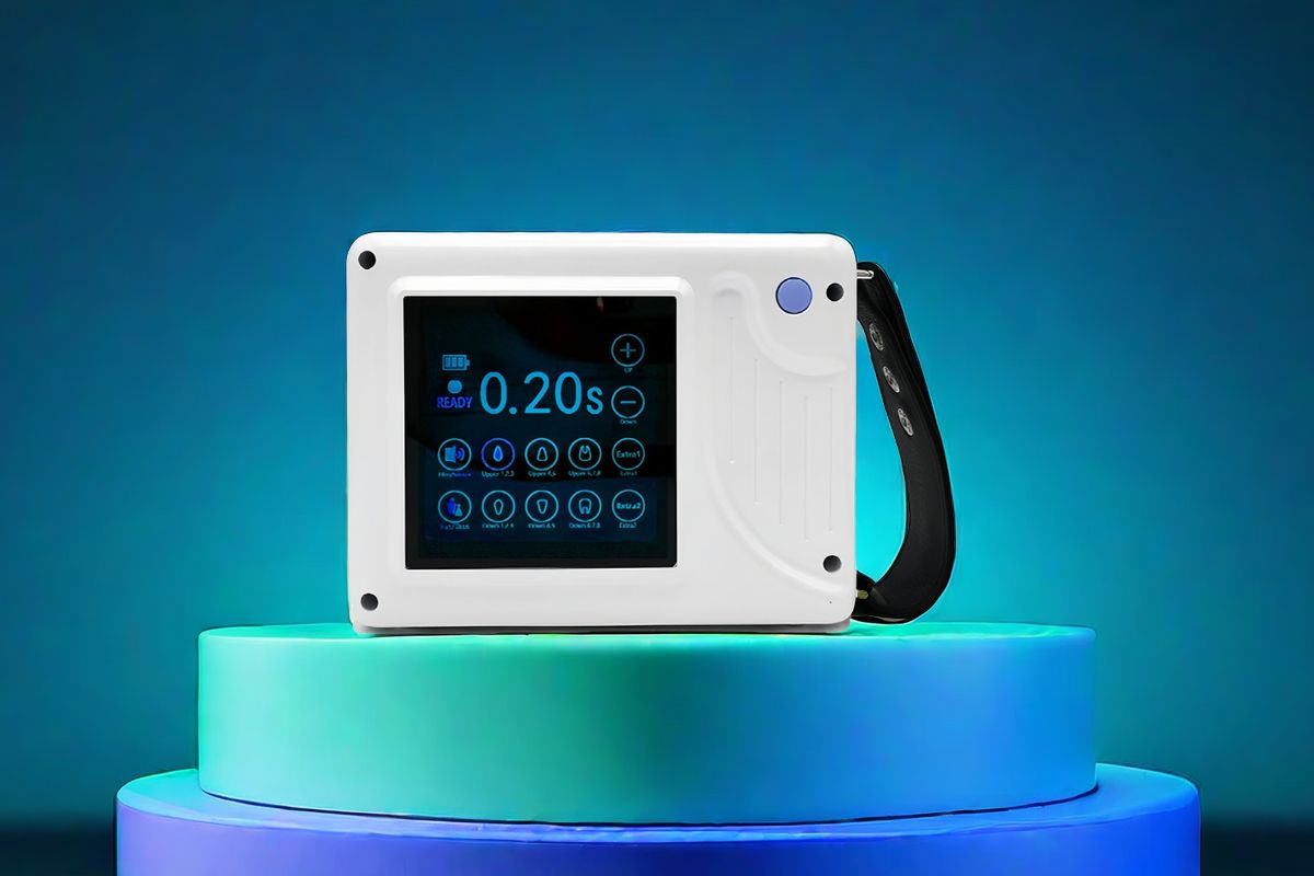
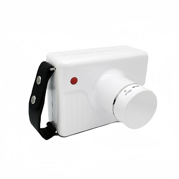
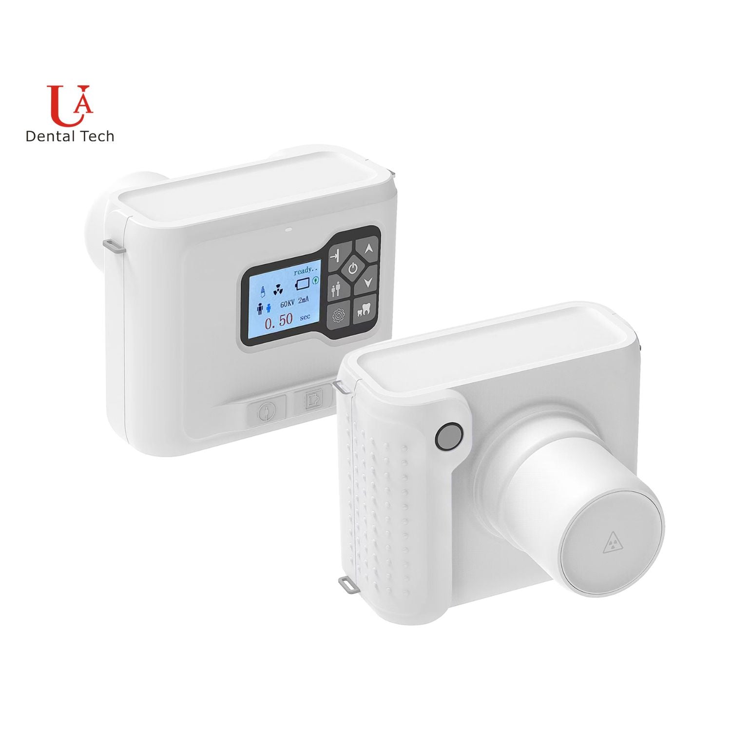
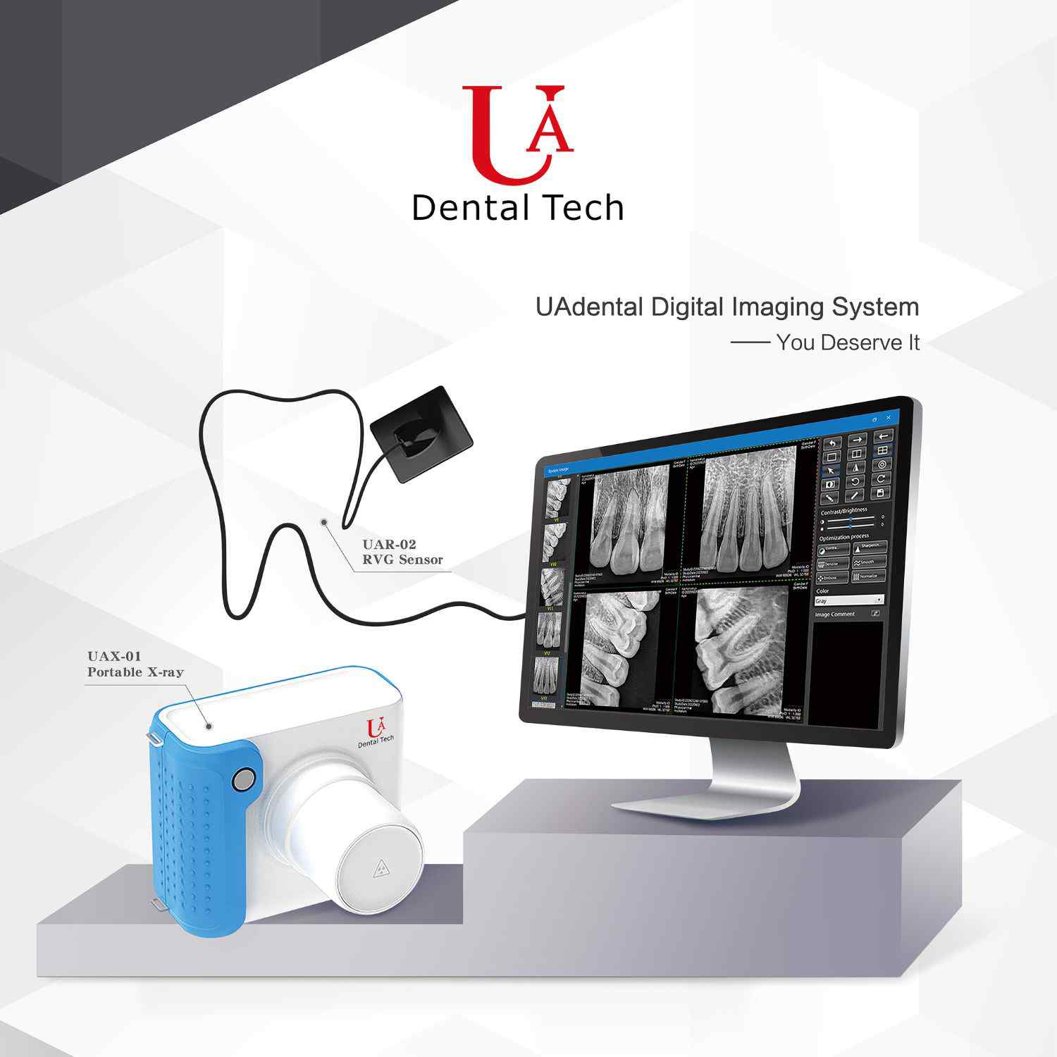
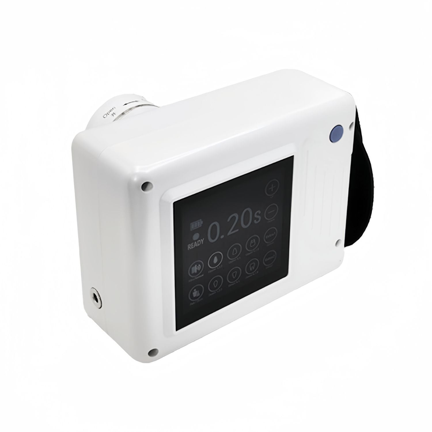
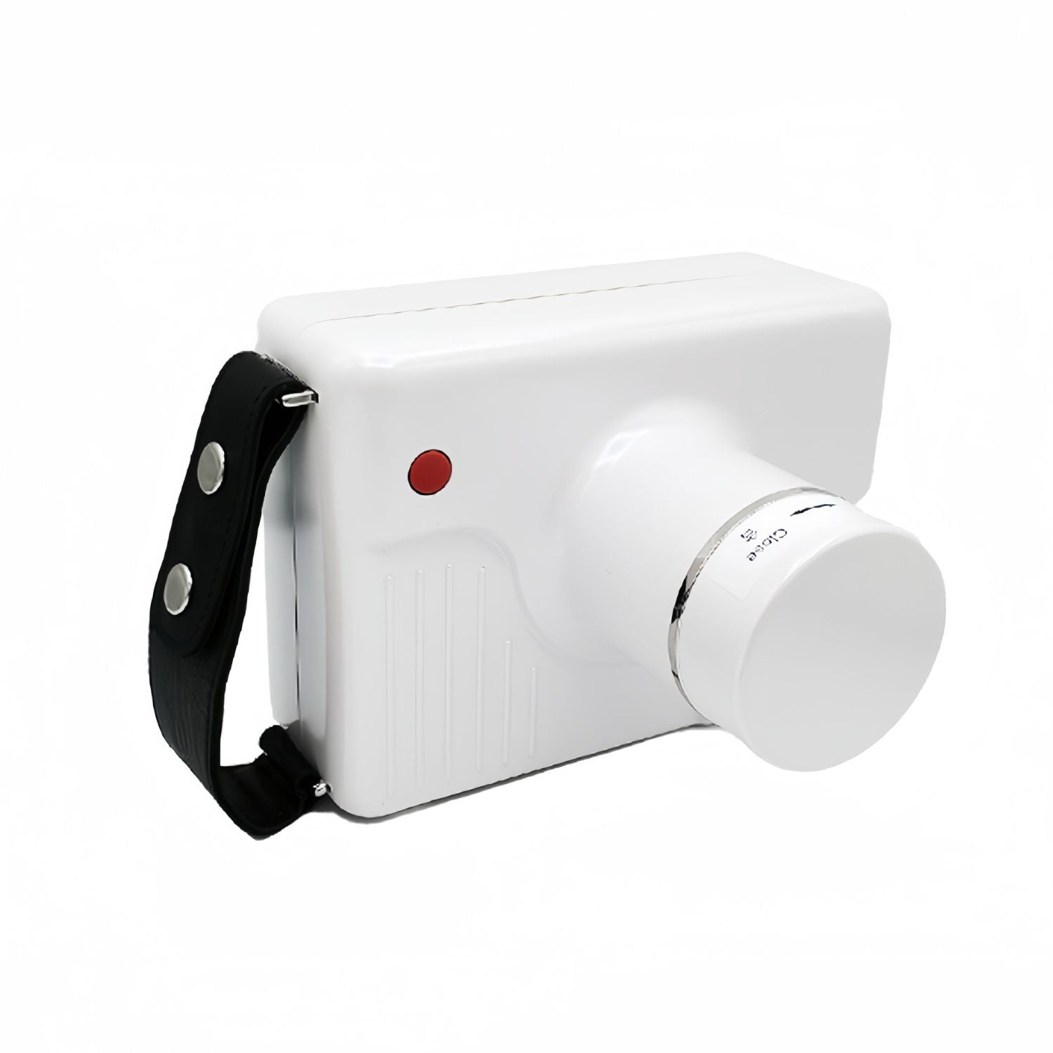
Dental X Ray Machine
The Dental X-ray Machine is a vital diagnostic tool used in dental clinics to capture detailed images of the teeth, gums, and surrounding bone structure.
This machine provides high-resolution radiographs that help dentists accurately diagnose and treat various dental conditions, such as cavities, infections, and bone abnormalities.
Equipped with advanced technology, the dental X-ray machine ensures minimal radiation exposure to patients while delivering clear and precise images, making it an essential component of modern dental care.
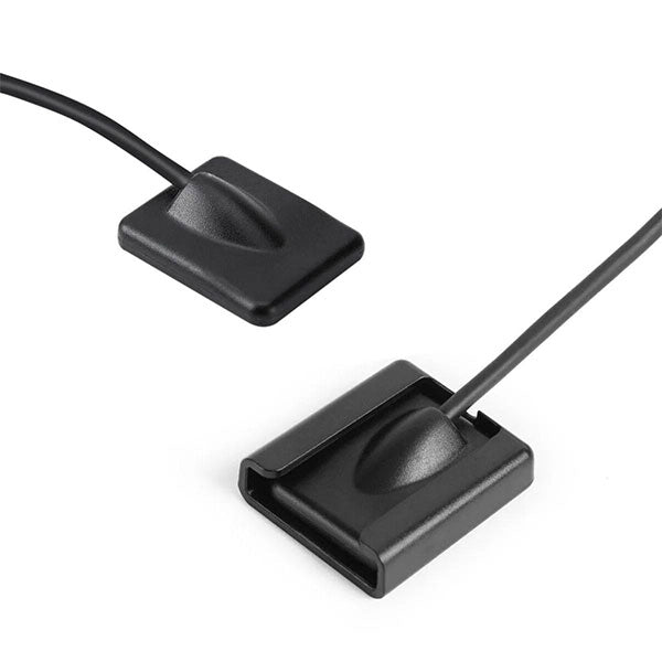
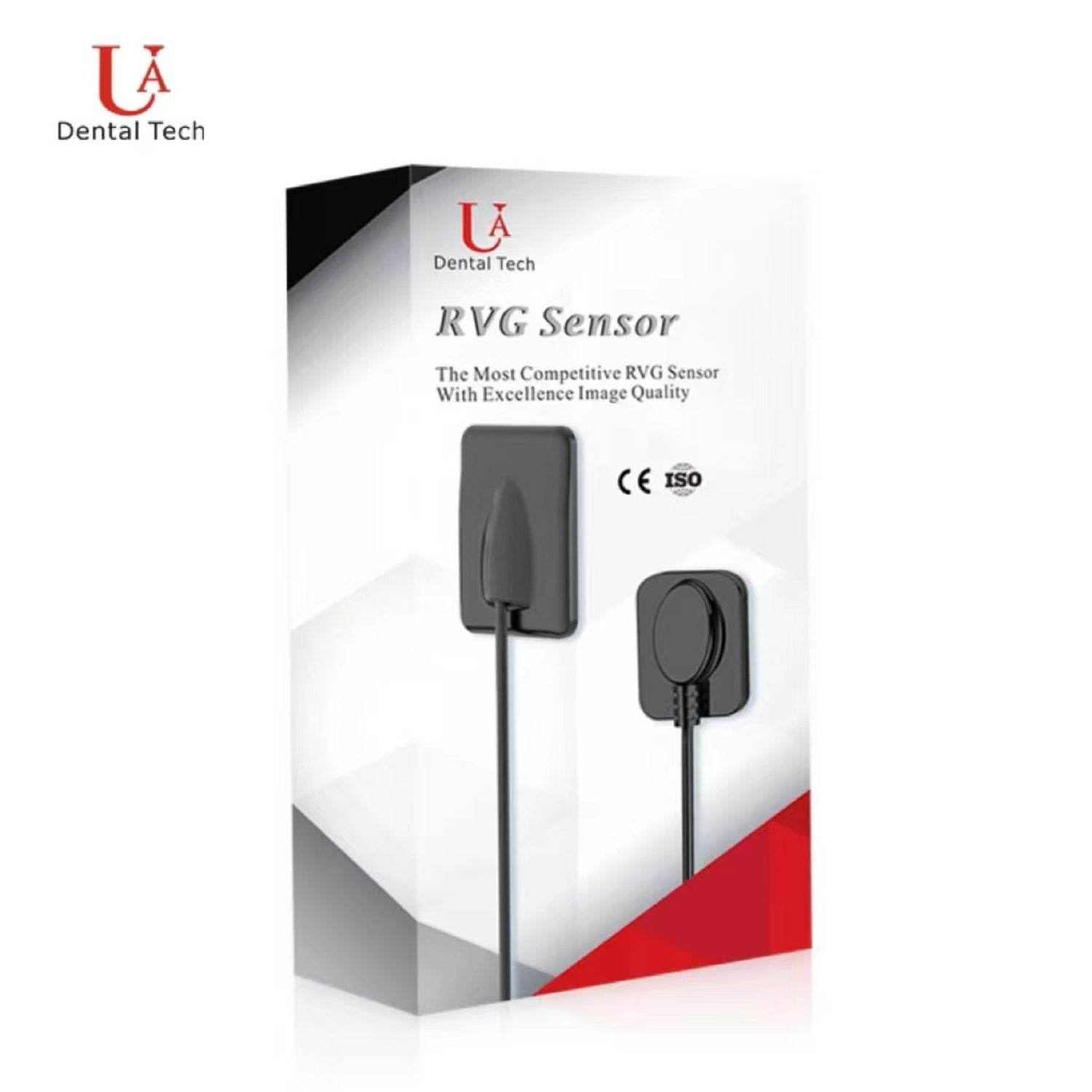
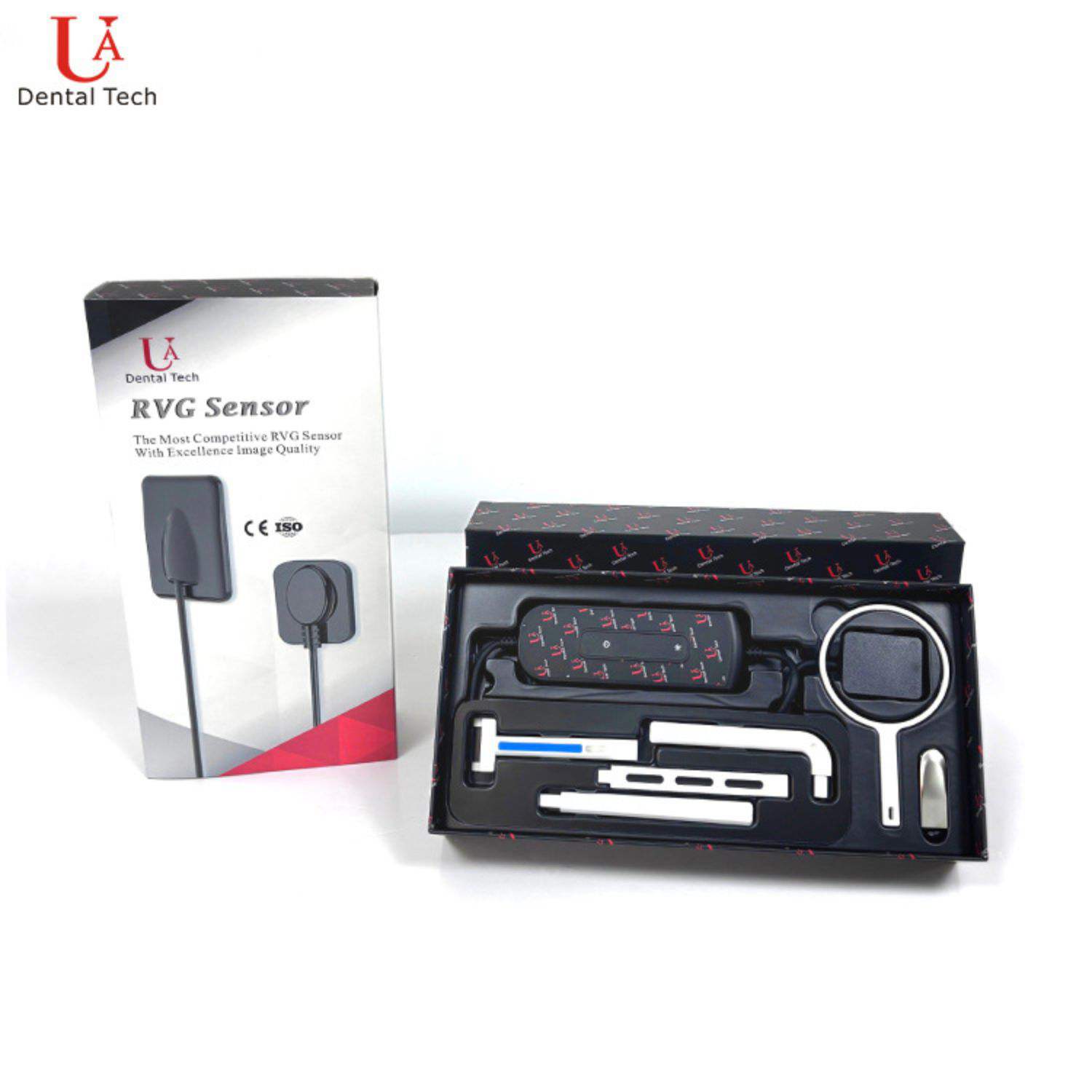
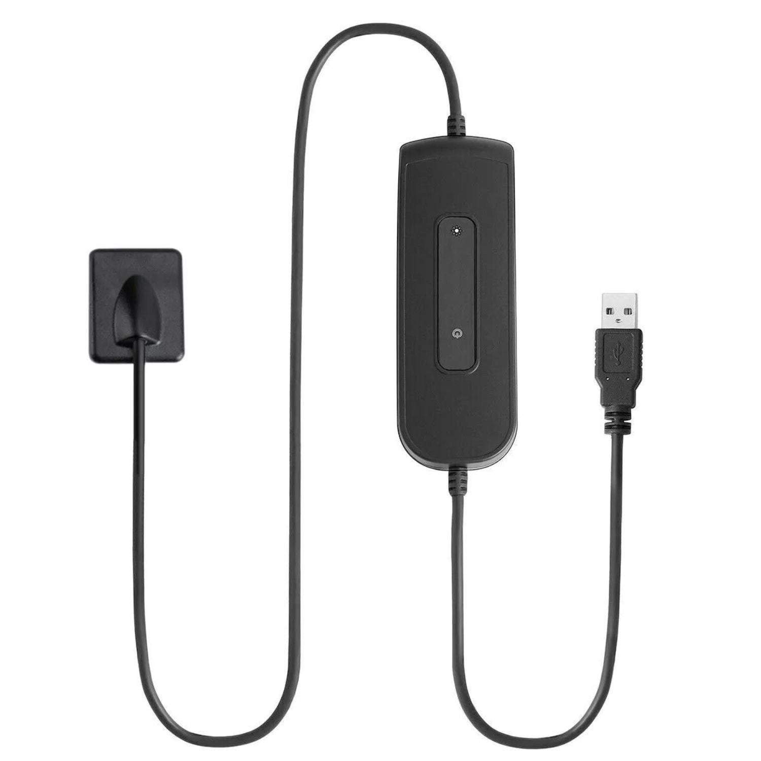
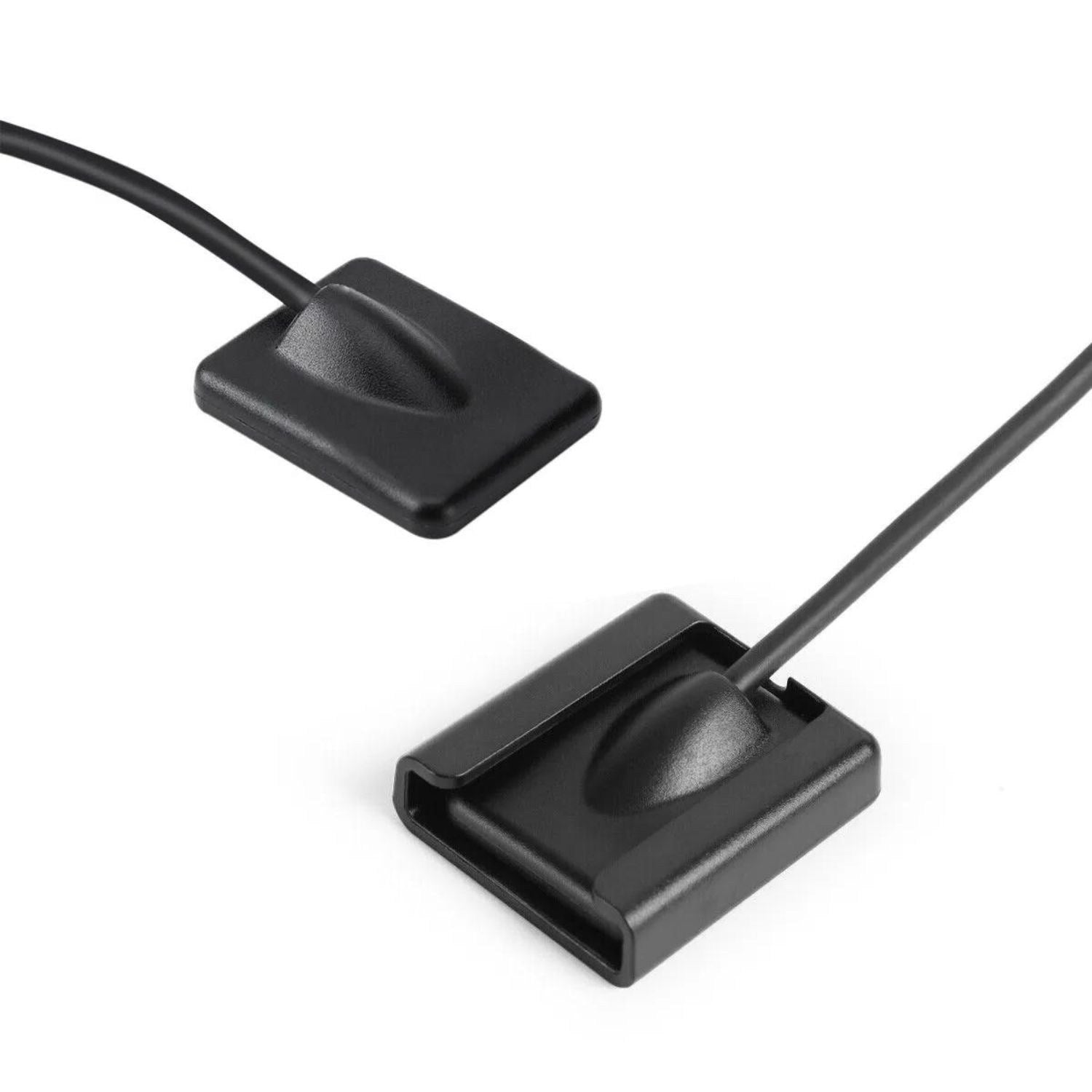
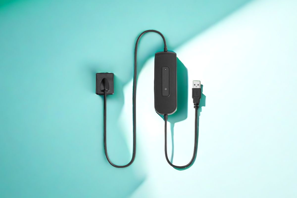
Dental Sensor
The Dental Sensor is a cutting-edge digital imaging device designed to capture high-quality intraoral X-rays.
It offers a significant improvement over traditional film X-rays by providing instant, clear images that can be viewed on a computer screen.
The compact and durable design of the dental sensor makes it easy to position in the patient's mouth, ensuring comfort and accuracy.
With its high sensitivity and ability to produce detailed images, the dental sensor aids in the early detection and treatment of dental issues, enhancing the efficiency and effectiveness of dental care.

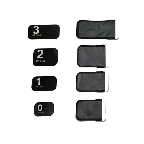
Phosphor Plates Dental
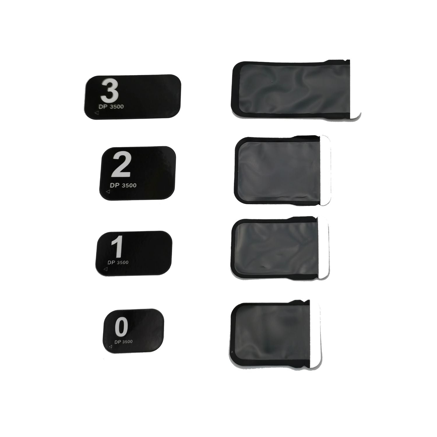
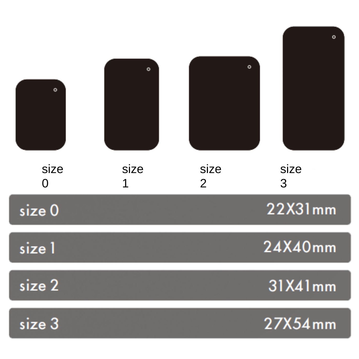
Phosphor Plates Dental are reusable imaging plates coated with a special phosphor material that captures X-ray images when exposed to radiation.
These plates are flexible and can be easily placed in the patient's mouth, offering a comfortable alternative to traditional film.
After exposure, the plates are scanned to produce high-resolution digital images.
Phosphor plates are known for their ability to capture detailed images with minimal radiation exposure, making them a popular choice in digital dental radiography.
Their reusable nature also makes them a cost-effective and environmentally friendly option.
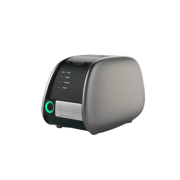
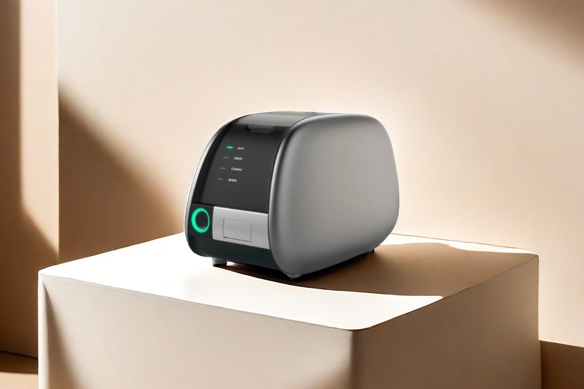
PSP Scanner
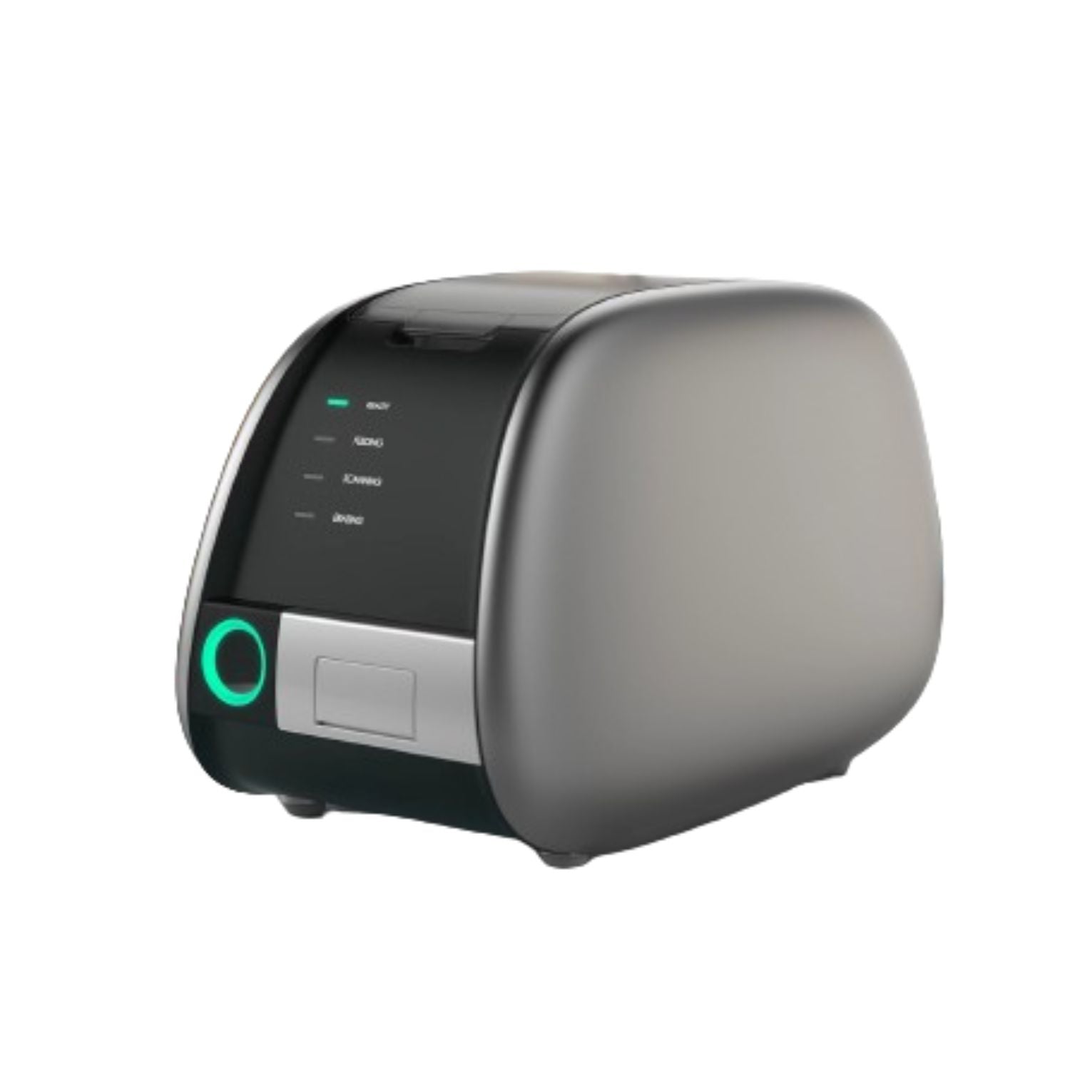
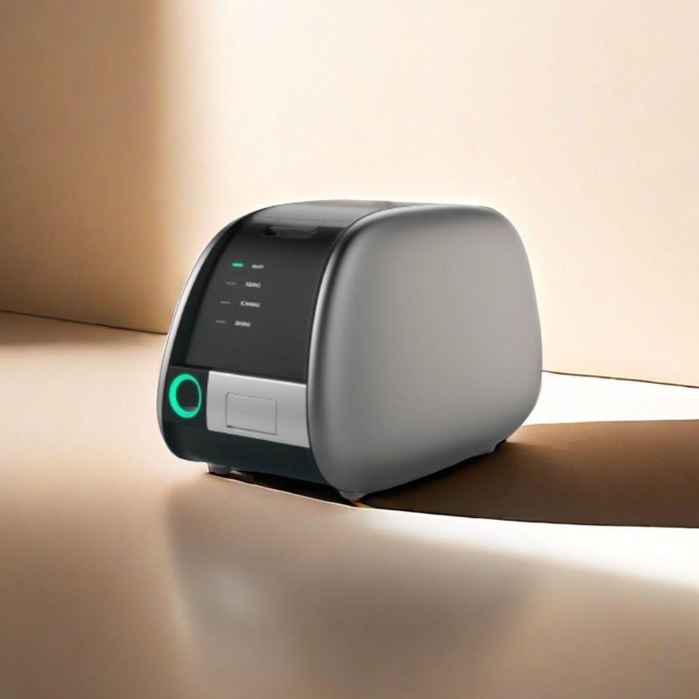
The PSP (Photostimulable Phosphor) Scanner is an essential device used to read and digitize images captured on phosphor plates.
After the plates are exposed to X-rays, they are inserted into the PSP scanner, which uses laser technology to extract and convert the stored energy into a digital image.
The resulting images are high-quality and can be easily viewed, stored, and shared electronically.
The PSP scanner streamlines the imaging process, reduces wait times for patients, and enhances diagnostic capabilities, making it a valuable addition to any dental practice.
FAQ
To obtain dental X-ray certification, enroll in an accredited dental radiography program offered by community colleges, technical schools, or online platforms.
Complete the required coursework and clinical training, then pass a certification exam such as the Radiation Health and Safety (RHS) exam by the Dental Assisting National Board (DANB).
Ensure you meet any additional state-specific requirements. Check with your state dental board and professional organizations like DANB for approved programs and specific certification details.
To read a dental X-ray, first familiarize yourself with normal dental anatomy and ensure the image quality is clear. Identify key structures such as teeth, bones, and soft tissues.
Look for common issues like cavities, bone loss, abscesses, and fractures. Correlate findings with the patient's symptoms and clinical examination, and consult with colleagues if needed.
Finally, document and communicate your findings clearly, and recommend appropriate treatment plans.
Obtaining X-ray certification as a dental assistant typically takes about 9-12 months.
This includes completing an accredited dental assisting program with specific courses in dental radiography, hands-on clinical training, and passing a certification exam like the Radiation Health and Safety (RHS) exam by the Dental Assisting National Board (DANB).
Some accelerated programs can be shorter, while part-time programs may take longer. Check state-specific requirements for any additional criteria.
Dental X-ray certifications typically do not have an expiration date and remain valid indefinitely.
However, dental assistants are often required to complete continuing education courses regularly to stay updated on best practices and regulatory changes.
State-specific requirements may vary, so it's essential to check with the state dental board for any specific renewal or continuing education requirements.
Disposing of a dental X-ray machine should be done in accordance with local regulations and environmental guidelines.
Contact regulatory authorities or hire professional disposal services specialized in medical equipment. Ensure proper decontamination and separation of reusable components.
Dispose of the machine through authorized channels, such as medical equipment disposal companies or recycling facilities, and keep documentation of the disposal process for accountability. Consider donation or resale if the machine is still functional.
Any question?
If we still haven't answered your question, you can contact us below and we will get back to you as soon as possible.

